Q1: Can different tissue types be loaded onto the same chip?
A: It is not recommended. Different tissue types often require distinct permeabilization conditions, and co-loading increases the risk of cross-contamination during the embedding process. Furthermore, once the tissue is mounted on the chip, the subsequent experimental steps must be performed immediately. Continuously mounting multiple tissue sections onto a single chip presents significant operational challenges and risks. Specifically, it is not feasible to mount sections from two different OCT blocks (processed in two separate cryostats) onto the same chip. Additionally, even when using two OCT blocks from the same cryostat, there is no guarantee of consistent data quality across the chip.
Q2: How does a gap between the tissue and the OCT affect the experiment?
A: OCT serves as an embedding medium that provides essential structural support for the tissue during sectioning and flattening. Without adequate OCT support, the tissue is highly prone to wrinkling and fragmentation during sectioning. Furthermore, areas where the tissue separates from the OCT tend to curl, which significantly complicates the mounting process.
Q3: Can bone samples be used for spatial transcriptomics?
A: Yes, but they carry significant risks. First, bone samples are heavily calcified and lack strong adhesion, making them prone to detaching from the slide (falling off); therefore, decalcification is usually required to enhance their adherence to the slide surface. Second, bone samples typically have low cellularity and low RNA content per cell. This often results in a low number of cells captured per spot and a low median number of genes detected.
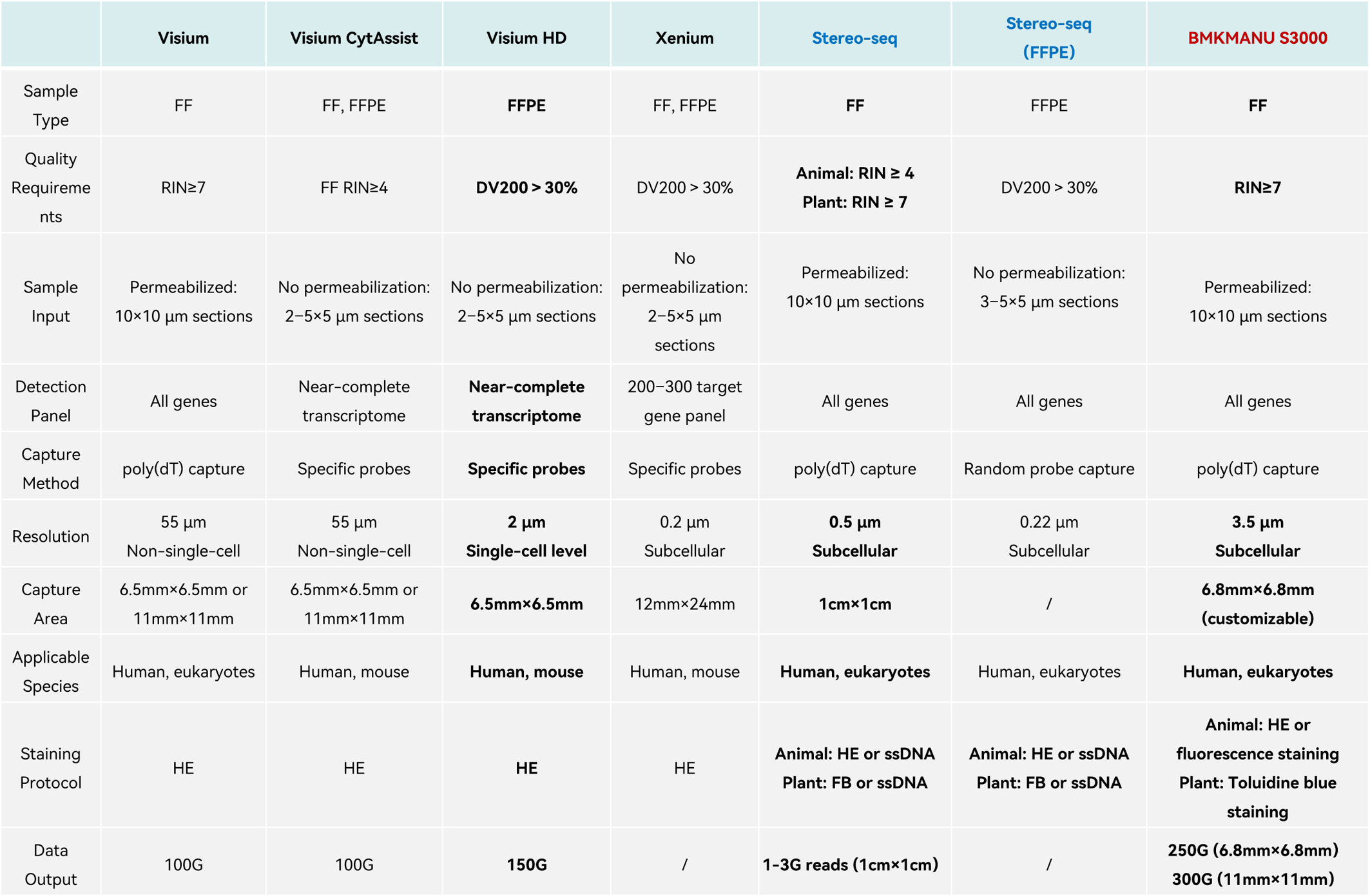
Q4: How do different spatial transcriptomics platforms compare?
Q5: What is a permeabilization test?
A: A permeabilization test is a pilot experiment designed to determine the optimal permeabilization time for a specific tissue section.
The test chip is loaded with nucleotide capture probes. After the tissue section is mounted, these probes capture mRNA molecules in situ. Fluorescently labeled nucleotides are then used for cDNA synthesis, allowing the optimal permeabilization time for the tissue to be rapidly assessed via fluorescence microscopy. Selecting the optimal permeabilization condition is crucial for efficient mRNA capture.
3 min permeabilization: The tissue exhibits uneven brightness across the same cortical layer, indicating insufficient permeabilization.
12 min permeabilization: The image shows clear details, uniform signal distribution, and maximum brightness.
24 min permeabilization: The signal intensity is weaker than that at 12 minutes.
Conclusion: Therefore, the optimal permeabilization time is 12 minutes.
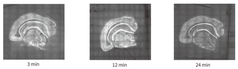
Figure. Fluorescence images of the permeabilization test.


 WeChat QR Code